Case Studies
Comparative Superiority
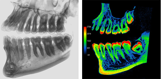
Conventional 2D images (Panorama / Dental) can only be evaluated from one settled angle.
On the contrary, 3D images provided by CBCT can be evaluated from different angles. Accurate and detailed information on inner structure can be obtained.
Case1: Root Fracture
The regular 2D x-ray image suggests a possible issue with an endodontic treatment. However, it is not sufficient for a clear diagnosis. The sagittal image of the 3D CBCT scan reveals a root facture.
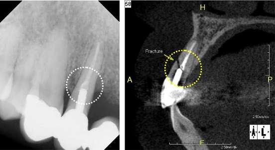
2D (Dental image) 3DMPR (Sagittal)
Case2: Maxillary impacted canine tooth
Slight shadow can be seen in the panoramic image. In the 3Dimage (MPR:Cpronal), the state of the eruption and the position of the maxillary impacted canine tooth can be accurately evaluated. Further detailed information can be obtained in the image of 3D VR.
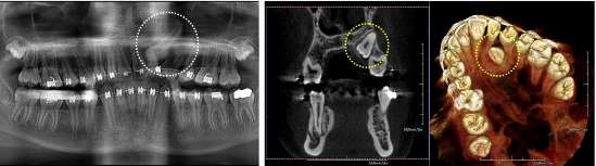
2D (Panoramic image) 3D (MPR:Coronal) 3D VR
Perio
The combination use of both 3D and multi-slice cone beam CT images provides a sharper understanding of the bony defects within relation to surrounding local anatomy.
In addition to helping the dentist plan for treatment options the patients can also view and become educated on any perio degenerated conditions for better acceptance of ensuing treatments.
Root abscess
Whatever the cause of root abscess be it from root canal failure , implants, etc... the PreXion3D scanner can evaluate and note the extent and involvement of surrounding anatomy and help plan the course of treatments to recommend. Liquified or purulent dense matter within abscess can be seen via the cone beam CT images.
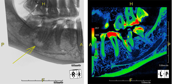
TMJ
The clarity and crisp detail of 3D imaging provide accurate assessment both of the position and condition and contour of the condyle including the upper ramus bone of the mandible.
Measurement tools applied reveal exact distances between the condyle and the bony fossa giving precise detail in aiding the design and/or positioning of corrective appliance.
Endo
PreXion scans aid in both pre endo treatments plus evaluation of post re-treatments of endodontic failures.
Information gathered from the cone beam scans can accurately determine if a re-treatment will be successful or if the tooth requires an extraction.
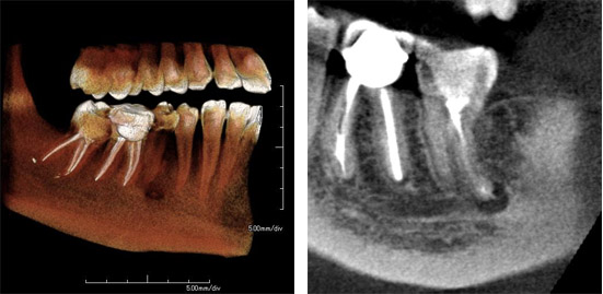
Molar
Both upper and lower molars more extensively evaluated for position, number of root canals and also the distance near the inferior alveolar canal if extractions are planned. Rotational and multi-slice features aid greatly and especially with deep or curved roots.